|
Tear of the lumbrical muscles
Definition
The lumbricals are small muscles of the hand that are not attached to the bones but to tendons. They are located between the tendons of the flexors and the extensor tendons of the finger at the palm of the hand (fig 1).
Their tearing, such as any muscular tearing, leads to brutal pain and a functional inability.
The pain is located in the palm. It can be spontaneous or occur while grabbing a hold (even an open hold) and at palpation.
It is possible that an oedema and/or a bruise appear.
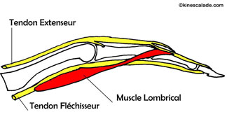
Fig 1: Situation of the lombrical muscle,
by M.Dufour, Anatomie de l'appareil locomoteur
Mechanism of the lesion
A one finger hold grabbed with a stretched finger while the others are bending, stretch the correspondent lumbrical muscle irreparably.
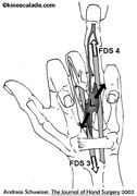
When the muscle is clinging to the flexor’s tendons of the middle and the ring fingers and one of these 2 fingers is maintained in extension while the other is stretched in flexing, the lumbrical is stretched between the 2 fingers (fig 2).
A muscle that is not correctly prepared cannot maintain such a load. On a one finger hold, risking appearing as Lapalisse, the body’s weight is entirely suspended on one (small) finger (fig 3).
 |
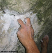 |
Fig 2: Strain that causes the lesion
of the lumbrical muscle with an one finger hold |
Fig 3: High-risk
one finger hold |
Prevention
Technique correction:
It is imperative to modify the grabbing technique of one finger holds by never separating the middle and the ring finger (fig 4).

Fig 4: Risk free grabbing of a one finger hold
Correction of the physical preparation:
It is imperative to warm up the muscle first; a well prepared muscle is less likely to tear.
An extensible muscle tears less: specific stretching exercises (see the page stretching videos) of ones muscles should be done systematically after each session.
Treatment
Stop the session and put some ice on it immediately. You also might want to try a compression (not really easy in that area).
Repeat ice treatment 3 to 4 times per day for 3 to 7 days. Do not apply anti-inflammatory gel.
A specific physical therapy combining ultrasound therapy, mobilization and stretching can help to optimize recovery.
Stop training for 10 to 30 days (depending on the importance of the lesion, determined by a sonogram). When you start training again do so progressively by strapping the middle finger together with the ring finger and without using one finger holds.
|